
Wrist Fractures
Jacksonville, FL
R. David Graham, MD is a fellowship-trained hand and upper extremity surgeon who treats wrist fractures as a core part of his practice. From simple distal radius fractures to complex, comminuted injuries in patients with osteoporosis, Dr. Graham brings subspecialty hand and upper extremity training to every case.


Understanding the Injury
What Happens When You Break Your Wrist
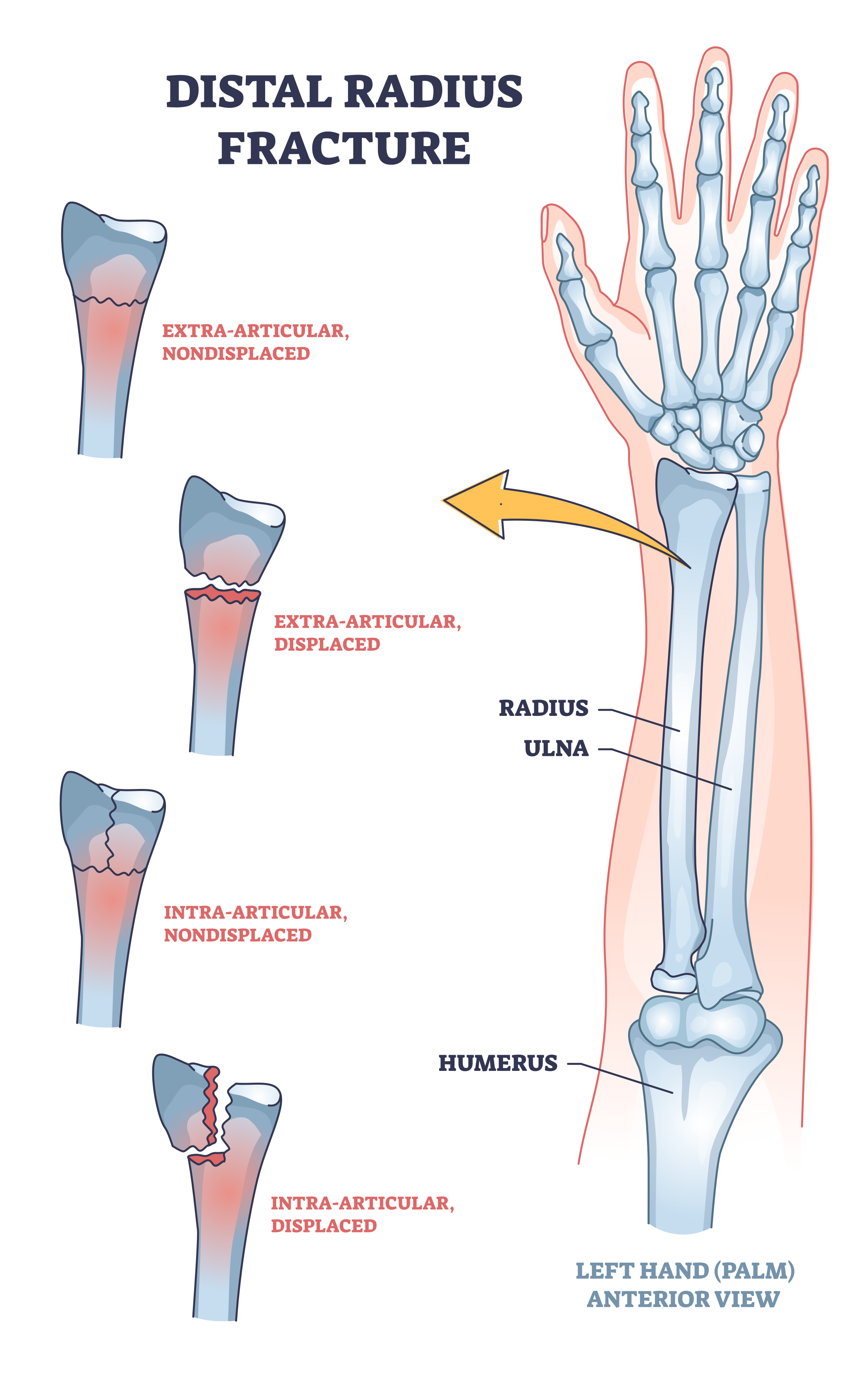
A wrist fracture is a break in one or more of the bones that make up the wrist joint — most commonly the distal radius, which is the larger of the two forearm bones and forms the primary load-bearing surface of the wrist. The distal radius is the most commonly broken bone in the body in adults over 50, and one of the most common fractures across all age groups.
The typical mechanism is a fall on an outstretched hand — instinctively extending the arm to break a fall. The force travels up through the palm and concentrates at the wrist, often fracturing the radius just above the joint. At higher energy levels — car accidents, sports impacts, falls from height — the fracture can be more complex, involving the joint surface, the ulna, the carpal bones, or surrounding ligaments.
The wrist is not simply a hinge. It is a precise, multi-plane joint on which the hand depends entirely for positioning, load transfer, and fine motor function. Getting the alignment right — and keeping it right through healing — is the central challenge of wrist fracture treatment. An incompletely reduced fracture or one that slips in a cast can result in a wrist that aches, stiffens, and loses strength for years.
This is why subspecialty focus and specialization matter. Decisions made in the first days after a wrist fracture determine long-term outcomes.
The distal radius — the most commonly fractured bone in the wrist — forms the primary joint surface. Its alignment determines how the wrist moves, how load travels through the joint, and whether arthritis develops years later.
The scaphoid (highlighted) is the second most commonly fractured wrist bone and is notoriously difficult to diagnose. It can feel like a sprain. Missed scaphoid fractures can lead to avascular necrosis and long-term wrist arthritis if left untreated.
Types of Wrist Fractures
Not All Broken Wrists Are the Same
The fracture pattern, bone involved, degree of displacement, and patient factors all influence treatment. Dr. Graham treats the full spectrum.
Distal Radius Fracture
The workhorse of wrist fracture surgery. The distal radius fractures at the metaphysis — just above the wrist joint — typically from a fall on an outstretched hand. It ranges from simple, minimally displaced injuries treatable with a cast, to severely comminuted fractures involving the joint surface requiring plate fixation.
Scaphoid Fracture
The scaphoid is the small boat-shaped bone on the thumb side of the wrist. It fractures when a fall drives the wrist into extreme extension. The injury can feel and look like a sprain — swelling is often minimal, bruising may be absent, and standard X-rays can miss the fracture entirely in the first days after injury.
Complex & Periarticular Fractures
Higher-energy mechanisms — car accidents, motorcycle crashes, falls from height, athletic impacts — can produce fracture-dislocations, fractures with associated ligament injuries, or multi-bone wrist injuries. These require careful assessment of both the osseous and ligamentous anatomy and often involve staged treatment.
Metacarpal & Phalangeal Fractures
Fractures of the metacarpals (the long bones in the palm) and phalanges (finger bones) are among the most common hand injuries. Boxer's fractures (fifth metacarpal neck from a punch) are the most recognizable, but any metacarpal or phalangeal fracture can cause lasting grip loss, rotational deformity, or stiffness if improperly treated. Rotational alignment is particularly critical — a subtle malrotation causes the fingers to scissor when making a fist.
Osteoporotic Wrist Fractures
In patients with osteoporosis — particularly women over 60 — even a low-energy fall from standing height can produce a severely comminuted distal radius fracture. The bone is too soft to hold a cast reduction reliably, and the fracture pattern is often more complex than in younger patients with similar mechanisms.
Distal Ulna Fractures
The ulna — the smaller forearm bone — often fractures alongside the distal radius, particularly the ulnar styloid. Isolated distal ulna fractures are less common but can destabilize the distal radioulnar joint (DRUJ), affecting forearm rotation and grip. Associated TFCC injuries are common.
Evaluation
Getting the Right Diagnosis
Wrist fracture evaluation begins with a careful examination and plain X-rays. For distal radius fractures, standard wrist views are usually sufficient to identify the fracture, characterize its pattern, and determine initial treatment.
CT scan is valuable when the fracture involves the joint surface (intra-articular fractures) and the degree of step-off or comminution needs to be precisely understood before surgical planning. CT is also essential for suspected scaphoid fractures when X-rays are inconclusive.
MRI is the most sensitive test for occult scaphoid fractures — fractures that are clinically suspected but not visible on X-ray or CT. It also identifies associated soft tissue injuries: TFCC tears, interosseous ligament injuries, and bone bruising.
If you think you may have broken your wrist, do not wait. Prompt imaging and evaluation matters — particularly for scaphoid fractures, where delays of even a few weeks substantially worsen outcomes.
Treatment
Surgical vs. Non-Surgical Treatment
Not every wrist fracture requires surgery. The decision depends on fracture pattern, alignment, patient age, activity level, and bone quality. Dr. Graham evaluates each fracture individually.
Many distal radius fractures — particularly in older, lower-demand patients or those with minimal displacement — can be treated with a cast after a closed reduction (realigning the bone by manipulation, without surgery). The wrist is immobilized for 6 weeks while the fracture heals.
The critical limitation of casting is that bone can shift during healing. Regular follow-up X-rays are essential in the first 2–3 weeks to confirm the fracture maintains acceptable alignment. If it does not, surgical fixation may still be required.
- Appropriate for minimally displaced or stable fractures
- Short arm or long arm cast, typically 4–6 weeks
- Serial X-rays to monitor alignment
- May require conversion to surgery if fracture displaces
- Hand therapy begins promptly after cast removal
ORIF — Open Reduction Internal Fixation — uses a plate and screws to hold the fracture in anatomic alignment while it heals. For displaced or unstable fractures, or those involving the joint surface, surgical fixation provides more reliable alignment, earlier mobilization, and better long-term function than casting alone.
Modern volar locking plates are low-profile, designed for the specific anatomy of the distal radius, and in most cases remain permanently without causing symptoms. Plate removal is not routinely recommended.
- Indicated for displaced, unstable, or intra-articular fractures
- Volar locking plate — designed for distal radius anatomy
- Allows earlier finger and wrist motion than casting
- Performed at Baptist Beaches Hospital — outpatient same day
- Hand therapy begins within 1–2 weeks of surgery
- Arthroscopic-assisted reduction available for select intra-articular fractures — direct visualization of the joint surface where it matters most
- Latest implant technology — Dr. Graham consults for orthopaedic industry
Anesthesia
ORIF of the distal radius is performed under a regional nerve block (arm block), which numbs the limb from the shoulder down. Depending on patient preference and health, Dr. Graham performs these with block alone, block plus MAC (monitored anesthesia care / light sedation), or general anesthesia. The procedure typically takes 45–75 minutes. Surgery is performed at Baptist Beaches Hospital or an affiliated surgery center — outpatient, home the same day.
Fracture Exposure & Reduction
An incision on the palm side (volar approach) of the wrist allows access to the fracture. The fracture fragments are carefully reduced — realigned to their anatomical position — under fluoroscopic (live X-ray) guidance. In complex intra-articular fractures, the joint surface is reconstructed fragment by fragment.
Arthroscopic Assistance (Select Cases)
For fractures involving the joint surface — particularly those with significant comminution or depression of the articular cartilage — Dr. Graham uses wrist arthroscopy as an adjunct to confirm intra-articular congruency. A small camera is introduced into the wrist joint, allowing direct visualization of the joint surface that fluoroscopy alone cannot provide. This ensures the cartilage is reconstructed as precisely as possible before the plate is applied.
Plate & Screw Fixation
A low-profile volar locking plate is applied to the radius and secured with locking screws that engage both the plate and the bone. This construct holds the fracture rigidly in alignment throughout healing. Fluoroscopy confirms screw length, plate position, and joint reduction before closure.
Closure & Recovery
The incision is closed in layers. A splint is applied for the first 1–2 weeks. Finger movement is encouraged immediately. Most patients begin formal hand therapy within 2 weeks of surgery to restore motion and strength. You go home the same day.
Revision & Complex Cases
When the First Surgery
Wasn't Enough
Wrist fractures that were treated elsewhere — surgically or non-surgically — sometimes fail to heal correctly, lose alignment, or leave patients with persistent pain and limited motion. When other orthopaedic surgeons encounter a difficult distal radius revision, they refer to Dr. Graham.
If your wrist was treated without surgery and continues to be painful or stiff — weeks, months, or even years later — you may be a candidate for malunion evaluation. A malunion occurs when a fracture heals in an incorrect position, altering wrist mechanics, limiting motion, and causing pain with activity. Malunion correction (osteotomy) can restore alignment and significantly improve function even long after the original injury.
Dr. Graham evaluates and treats the full spectrum of revision wrist surgery — including hardware failure, infection, malunion, nonunion, and complex reconstruction after failed primary treatment — whether the original surgery was performed here or elsewhere. Second opinions are welcome.
Recovery
What to Expect After Wrist Fracture Treatment
Recovery timelines vary by fracture severity, treatment type, patient age, and bone quality. This represents a typical surgical recovery for a distal radius ORIF.
Home the Same Day
Surgery is outpatient. You go home in a splint with fingers free to move. Elevate the hand above heart level to minimize swelling. Pain is typically well-controlled with oral medications. Dr. Graham's team will contact you to check in.
wks
Splint, Elevation, Finger Motion
The splint protects the repair while the incision heals. Finger movement is encouraged — open and close the hand regularly to prevent stiffness and reduce swelling. Sutures are removed at 10–14 days. You are seen in clinic for a wound check and X-ray confirmation of the reduction.
wks
Hand Therapy Begins — Wrist Motion Returns
Formal hand therapy begins, focusing on wrist flexion, extension, forearm rotation, and edema management. A removable thermoplastic splint replaces the post-op dressing. Most patients achieve meaningful wrist motion during this phase. Light grip activities are introduced progressively.
wks
Strengthening Phase
Fracture healing is confirmed on X-ray. Resistive exercises and grip strengthening begin. Most patients are performing the majority of daily activities without significant restriction. Return to driving typically possible by 6–8 weeks depending on which wrist was injured and type of transmission.
mos
Return to Near Pre-Injury Levels
The majority of active patients — athletes, working adults, recreational sportspeople — return to near pre-injury levels of function. Return to heavy labor or sport varies from 3–6 months depending on fracture complexity and individual recovery. Final grip strength typically reaches 80–90% of the opposite side. Some residual stiffness or mild weather-related aching is common and continues to improve. For most patients, the wrist does not define their limitations after this point.
Complex Cases
Wrist Fractures Require More Experience in Certain Patients
Subspecialty training and experience matter most when the case is not straightforward. Dr. Graham's practice sees all of these regularly.
Patients with Osteoporosis
Osteoporotic fractures are more comminuted, less amenable to casting, and require implants designed for poor bone quality. Locking volar plates provide stable fixation in osteoporotic bone and allow earlier mobilization — critical in older patients for whom prolonged immobilization carries its own risks. A distal radius fracture in an osteoporotic patient is also a sentinel event signaling the need for bone health evaluation and treatment.
Active & Working Adults
Patients with physical jobs, active lifestyles, or sport demands cannot afford months in a cast with an uncertain alignment outcome. Surgical fixation with early mobilization — hand therapy starting at 2 weeks — consistently produces faster return to function and activity in this group. Dr. Graham takes return-to-work and return-to-sport timelines seriously at the initial consultation.
Athletes
Mickey, Dr. Graham's certified athletic trainer with MLB experience from the Philadelphia Phillies organization, helps bridge the gap between surgical recovery and sport-specific rehabilitation. Athletes benefit from a team that understands performance demands, not just functional milestones. Scaphoid fractures in young athletes — often misdiagnosed as sprains — are a particular area of focus.
Why Experience Matters
When You Break Your Wrist,
Experience Is Everything
Wrist fractures are not all created equal, and neither are the surgeons who treat them. A fellowship-trained hand and upper extremity surgeon who operates on wrist fractures regularly brings a different level of pattern recognition, implant familiarity, and intraoperative decision-making than a general orthopaedic surgeon who sees one every few weeks.
R. David Graham, MD treats wrist fractures as a core focus of his subspecialty practice. Experience matters because fracture patterns vary widely, complications are recognizable earlier when you have seen the full range of them, and implant selection and surgical technique become more refined with repetition.
He also consults for orthopaedic industry companies — which means the newest plating systems and fixation techniques reach his patients as soon as the evidence supports their use. That is a meaningful advantage in a field where implant technology has advanced substantially in the last decade.
One detail patients consistently notice: the incision is smaller than they expected. Dr. Graham performs wrist ORIF through a minimal volar exposure — precise dissection rather than a large opening. The scar heals to a fine line that most patients say they can barely find months later. Pauline H., 71 years old with severe osteoporosis and a four-part fracture, noted "essentially no scar" at two months.
For most active patients — recreational athletes, working adults, people with physical hobbies — the goal is a return to near pre-injury levels of function. That is the expectation Dr. Graham sets at the first visit, and the outcome the majority of his patients achieve.
Clinical Results
Before & After
X-ray imaging and surgical photographs shared with patient consent. The incision speaks for itself.
The Incision
Nobody Does It Through a Smaller Incision
Precise, minimal dissection. The wrist ORIF incision Dr. Graham uses is deliberately small — enough for the anatomy, nothing extra. At two months, most patients say they can barely find it. Photos available at consultation.
pending
pending
pending
All images shared with written patient consent. X-rays de-identified.
Patient Experiences
In Their Own Words
“Everything was fully explained to me. Dr. Graham called me at home the evening of my surgery to see how I was doing. Ten weeks post surgery my wrist is back to normal.”
“Dr. Graham did an excellent job removing a plate in my wrist and doing a Carpal tunnel release. His bedside humor and surgical skills are just what I needed!!”
“Had an accident, needed to see surgeon for fractured wrist. Dr Graham was available and able to perform surgery soon thereafter. Three months later I am back to my normal routine.”
Common Questions